ScienceDaily (Mar. 9, 2009) — We usually think of fish as a "heart-healthy" food. Now fish are helping researchers better understand how heart disease develops in studies that could lead to new drugs to slow disease and prevent heart attacks.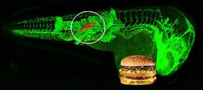
Scientists at the University of California, San Diego School of Medicine have done to laboratory zebrafish exactly what many people still do to themselves – added excess cholesterol to their diet. Because young zebrafish are transparent, researchers were able to see – literally – the development of plaques in the zebrafish blood vessels.
The study led by Yury I. Miller, MD, PhD, associate professor of medicine at UC San Diego School of Medicine will be published online March 5th in advance of print in the April issue of Circulation Research, published by the American Heart Association.
"The use of this transparent zebrafish model is a promising method to screen for new drugs and cardiovascular imaging agents," said Miller.
Atherosclerosis is a process of thickening and hardening of the artery walls as a result of fat deposits and inflammation. Risk factors for atherosclerosis include high levels of "bad" cholesterol, high blood pressure (or hypertension), smoking, diabetes and a family history of the disease – all of which can lead to heart attack or stroke.
Extreme hyperlipidemia, or the presence of excess fat and cholesterol molecules in the bloodstream, has been induced in mice and rabbits in the past, but microscopic examination of plaque build-up was only possible post-mortem.
Miller and colleagues fed a high-cholesterol diet (HCD) to zebrafish, supplementing the HCD with a red fluorescent lipid. They also used fish with endothelial cells – the thin layer of cells that line the interior surface of blood vessels – that were tagged with green fluorescent protein. Fish macrophages were tagged with red fluorescent protein, illuminating these immune cells which regulate chronic inflammation and indicate the development of atherosclerosis.
"Because zebrafish are transparent for the first 30 days of life, we can see in the living fish that the blood vessels glow green, while the fat deposits in vascular plaques are red," said Miller. He added that, interestingly, the zebrafish on a high-cholesterol diet did grow little fat fish stomachs.
The scientists used confocal microscopy, able to detect the fluorescent cells, in order to monitor vascular lipid accumulation and view the thickening of the endothelial lining in the living zebrafish. In other experiments, zebrafish in which the macrophages expressed red fluorescent protein were given HCD. This resulted in the pathologic accumulation of fluorescent macrophages along the endothelial cells of the vascular wall, as happens in human atherosclerotic plaques.
To explore the potential of zebrafish for atherosclerosis-related drug screening, the researchers administered the drug ezetimibe by adding it to the fish tank water. Ezetimibe is a medication marketed as Zetia, used to lower plasma cholesterol levels by lowering cholesterol absorption in the intestine. After treating the HCD-fed zebrafish with ezetimibe, the scientists were able to literally see that the drug significantly diminished the thickening of the vascular wall and improved its barrier function.
"Many researchers and clinicians agree that the treatment of atherosclerosis must begin at the earliest possible stage – the fatty streak," said Miller. "By feeding HCD to zebrafish, we were able to reproduce many of the processes involved in early atherogenesis. Our results suggest that this new model is suitable for studying inflammatory processes that occur in the early development of the disease, by looking at the function of vascular cells and lipid deposits in a live animal."
Co-first authors of the study are Konstantin Stoletov, UCSD Department of Pathology, and Longhou Fang, UCSD Department of Medicine. Other contributors include Soo-Ho Choi, Karsten Hartvigsen, Lotte F. Hansen, Jennifer Pattison, Joseph Juliano, Elizabeth R. Miller, Felicidad Almazan and Joseph L. Witztum, UCSD Department of Medicine; Chris Hall and Phil Crosier, Department of Molecular Medicine and Pathology, University of Auckland, New Zealand; and Richard L. Klemke, UCSD Department of Pathology. The study was a collaborative effort of the Miller and Klemke laboratories.
Funding for the study was provided by the National Institutes of Health and the Leducq Fondation.
Source: ScienceDaily


